Case Report: Carotid body paraganglioma in a 20-year-old woman: multimodal imaging, vessel-preserving surgical excision, and long-term clinical follow-up - Summary - MDSpire
Advertisement
Case Report: Carotid body paraganglioma in a 20-year-old woman: multimodal imaging, vessel-preserving surgical excision, and long-term clinical follow-up
To report a case of carotid body paraganglioma in a young woman, highlighting the diagnostic imaging and surgical approach in a case report format.
Approach:
Patient Presentation: A 20-year-old woman presented with a painless left lateral neck mass that had been present for over 5 years.
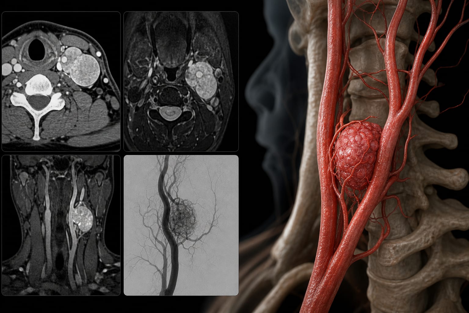
Diagnostic Imaging: Multimodal imaging including ultrasonography, CTA, MRI, and DSA was utilized for diagnosis and surgical planning.
Surgical Intervention: Surgical excision was performed with a focus on preserving the carotid arteries and surrounding nerves.
Follow-Up: Postoperative follow-up included ultrasonography and clinical assessment for recurrence.
Key Findings:
The tumor was hypervascular and involved the carotid bifurcation.
Histopathology confirmed the diagnosis of carotid body paraganglioma.
Surgical excision was grossly complete with preservation of major vessels and nerves.
Interpretation:
The case demonstrates the effectiveness of multimodal imaging in diagnosing carotid body paraganglioma and the feasibility of vessel-preserving surgical excision.
Limitations:
No formal microscopic margin status was specified in the pathology report.
Balloon occlusion testing, biochemical catecholamine work-up, functional imaging, and genetic testing were not documented.
Conclusion:
The case illustrates the management of carotid body paraganglioma in a young patient with follow-up outcomes.