To report a case of reactive lymphoid hyperplasia at the eustachian tube orifice and discuss its clinical characteristics, diagnostic challenges, and management.
Approach:
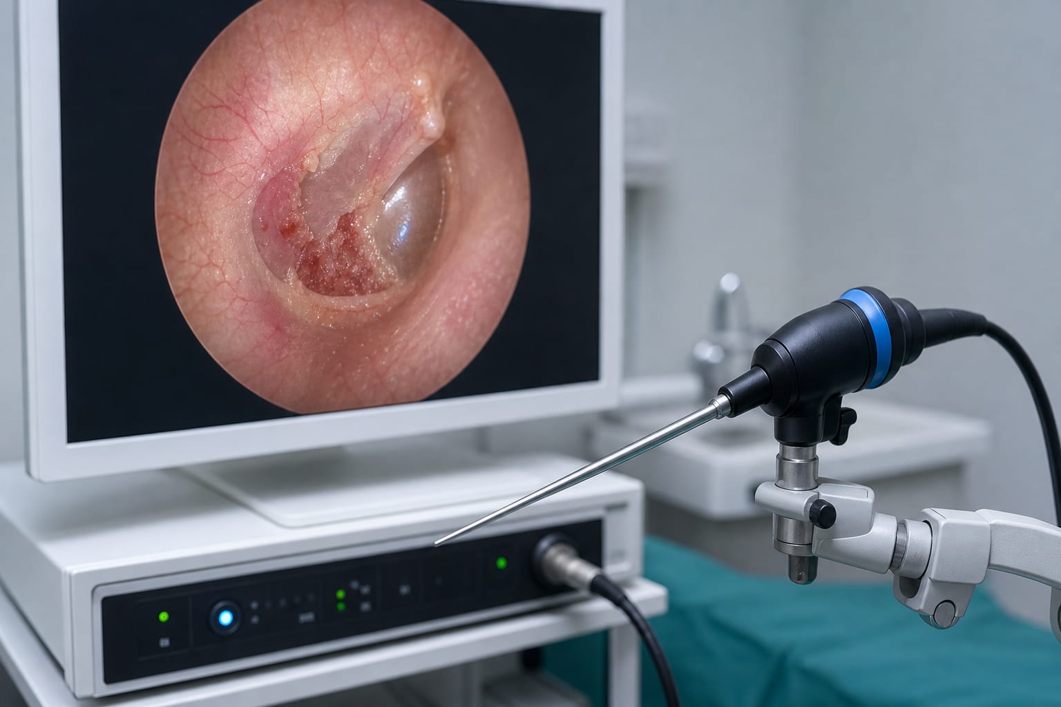
Patient Presentation: A 58-year-old male presented with persistent right-sided aural fullness and hearing loss, initially diagnosed with otitis media with effusion.
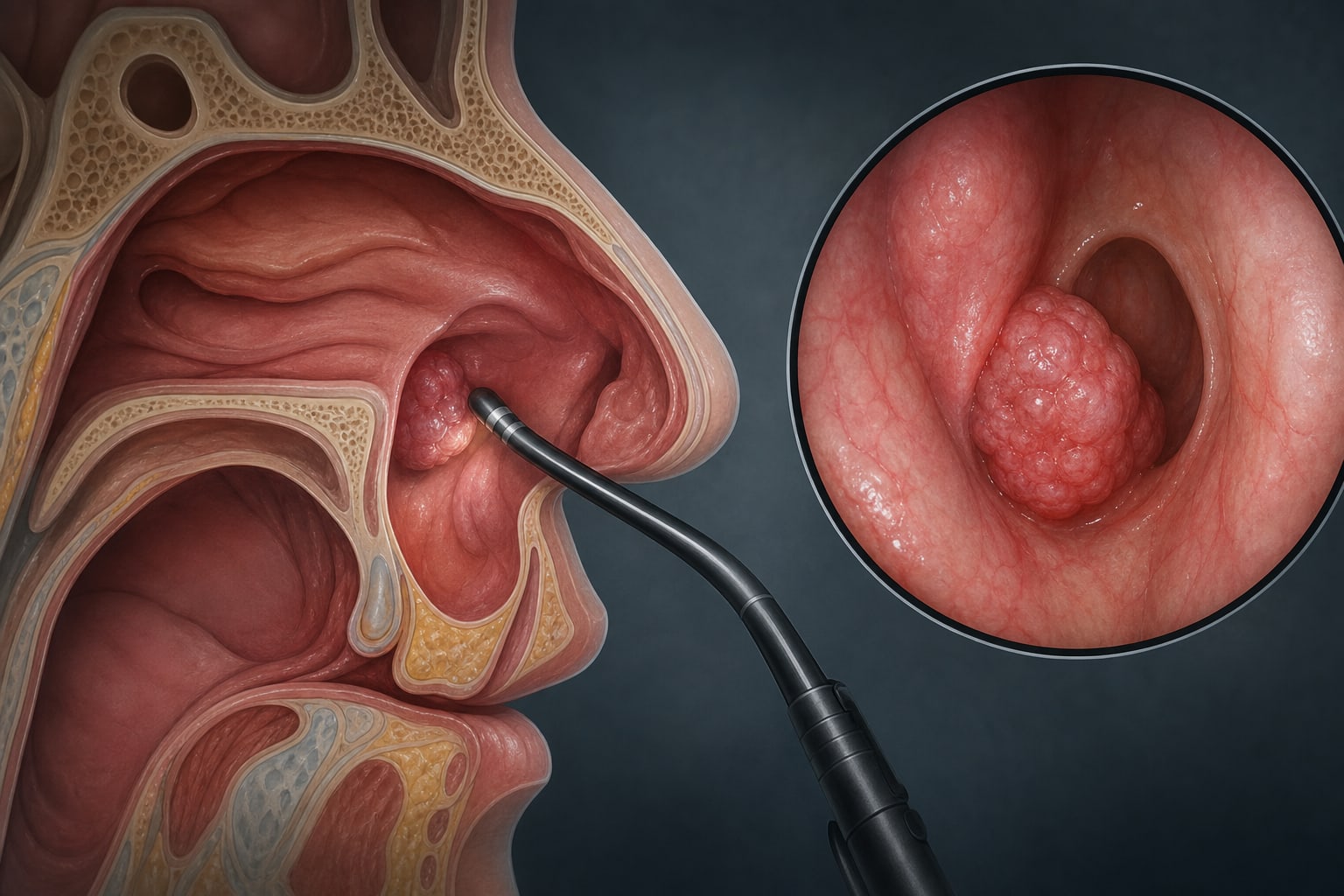
Diagnostic Evaluation: Nasal endoscopy revealed a neoplastic lesion at the right eustachian tube orifice, and biopsy suggested lymphoid hyperplasia.
Surgical Management: Navigated endoscopic resection of the mass was performed, confirming benign lymphoid hyperplasia through histopathological examination.
Follow-Up: Postoperative improvement was noted, with complete resolution of symptoms and no recurrence at 6-month follow-up.
Key Findings:
Reactive lymphoid hyperplasia is a rare benign lesion at the eustachian tube orifice, as demonstrated in this case.
Benign lesions can be misdiagnosed as malignant due to overlapping clinical and imaging features, as seen in the patient's presentation.
Histopathological confirmation was essential for accurate diagnosis in this case.
Interpretation:
This case emphasizes the importance of comprehensive evaluation and pathological confirmation in diagnosing lesions at the eustachian tube orifice.
Limitations:
The rarity of benign lymphoproliferative lesions at the eustachian tube orifice may limit the generalizability of this case.
Follow-up MRI was not performed postoperatively due to the resolution of symptoms, which may affect long-term assessment.
Conclusion:
This case provides practical insights for the diagnosis and treatment of rare lesions at the eustachian tube orifice.
Pain reprocessing therapy produced small but measurable reductions in cross-modal sensory amplification, pointing to a broader neurological signature of chronic back pain than previously recognized.