Incisura groove depth is associated with reduced rotational fibular displacement after syndesmotic injury: cadaveric validation of an automated three-dimensional incisura morphometric pipeline - Summary - MDSpire
Advertisement
Incisura groove depth is associated with reduced rotational fibular displacement after syndesmotic injury: cadaveric validation of an automated three-dimensional incisura morphometric pipeline
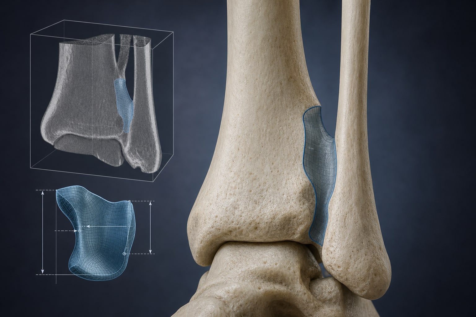
To develop and validate an automated three-dimensional morphometric toolbox for the tibial incisura and to test its outputs in a controlled cadaveric model of syndesmotic injury.
Approach:
Toolbox Development: An automated MATLAB toolbox was created to compute eight three-dimensional morphometric parameters of the incisura from CT-derived data.
Evaluation: The toolbox was evaluated on 34 cadaveric tibiae for reproducibility and bilateral reliability.
Cadaveric Protocol: Outputs were correlated with fibular displacement magnitude from paired six-degree-of-freedom kinematics after syndesmotic sectioning.
Key Findings:
All eight toolbox outputs demonstrated reproducibility across repeat CT acquisitions.
Bilateral reliability was good for all outputs, with seven exceeding ICC of 0.80.
Four of the six outputs tested showed significant negative association with external rotation magnitude: mean groove depth (r = −0.48, p = 0.004), maximum groove depth (r = −0.46, p = 0.006), conventional single-level incisura floor depth (r = −0.44, p = 0.010), and total groove volume (r = −0.37, p = 0.030).
The first principal component explained 85.4% of variance and was negatively associated with external rotation (r = −0.42, p = 0.015).
Interpretation:
The incisura metrics toolbox provides a reliable method for three-dimensional morphometric characterization of the tibial incisura, indicating that larger osseous constraint envelopes are associated with reduced rotational fibular displacement.
Limitations:
The study was conducted on cadaveric specimens, which may not fully replicate in vivo conditions, and the sample size was limited to 34 cadaveric tibiae, potentially affecting the generalizability of the findings.
Conclusion:
The study validates a method for assessing incisura morphology, suggesting its potential role in future diagnostic and predictive injury models.