To evaluate the effectiveness of in vivo confocal microscopy (IVCM) and anterior segment optical coherence tomography (AS-OCT) in diagnosing and monitoring infectious keratitis (IK).
Approach:
Key Findings:
OCT provides non-contact, high magnification images but is limited by back shadowing in dense infiltrates.
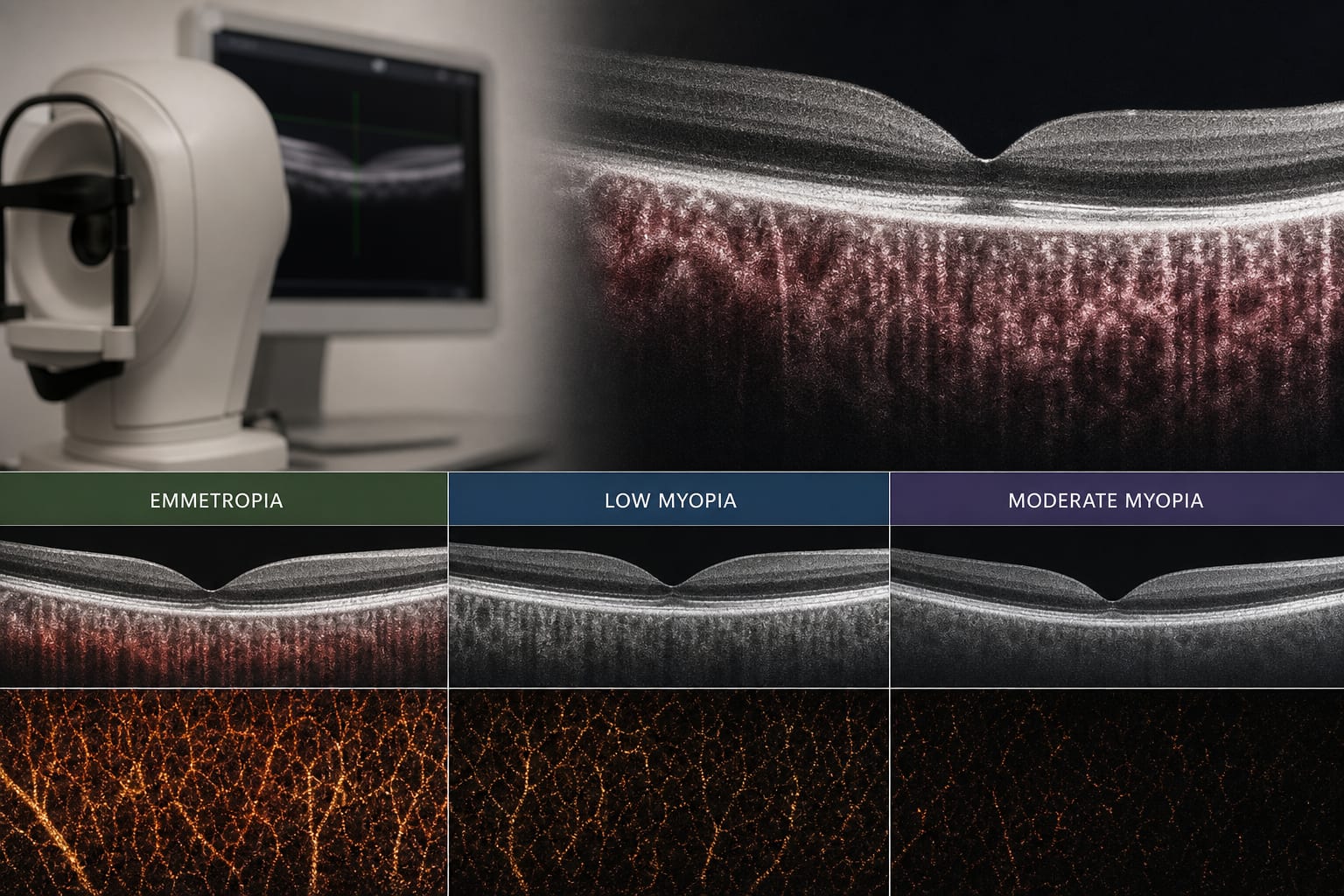
IVCM allows for cellular-level examination and can visualize invading organisms with high sensitivity and specificity.
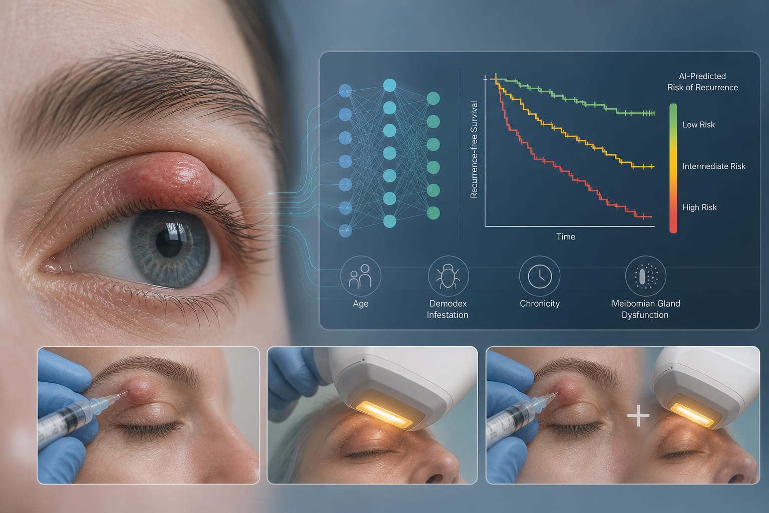
Deep learning models show promise for IK diagnosis but face challenges such as the need for large datasets and misclassification bias.
Interpretation:
Remove unsupported conclusions.
Limitations:
The study was limited to a single center and a small sample size of 35 patients.
The reliance on imaging quality and the need for experienced interpretation skills may affect outcomes.