To characterize the clinical, radiological, and histopathological features of Kimura disease (KD) presenting in atypical anatomical sites, particularly in the lower extremities.
Approach:
Key Findings:
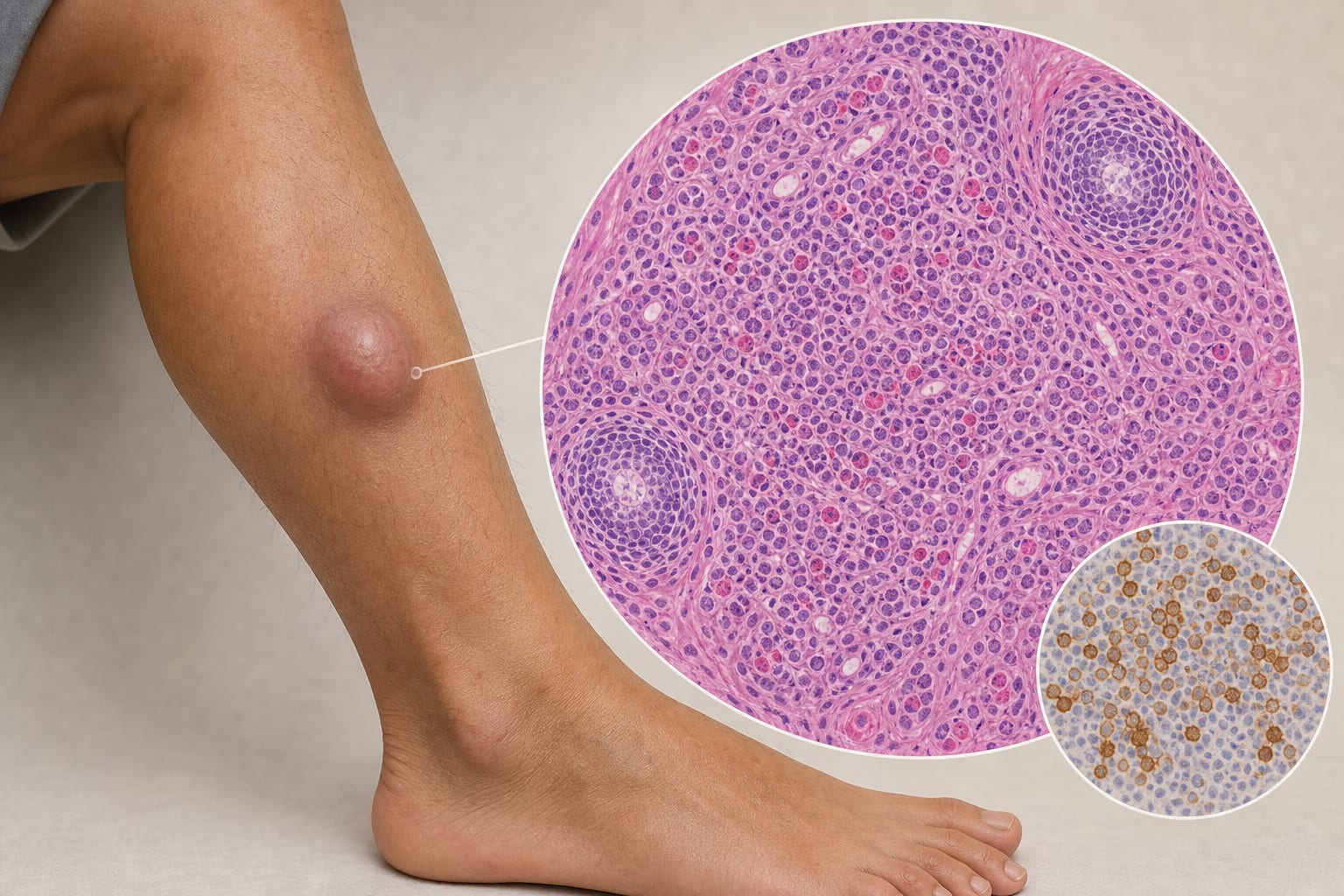
The patient presented with a spindle-shaped mass at the anterior border of the right tibia, measuring approximately 2 cm.
Imaging studies revealed a solid, well-circumscribed mass with internal vascular proliferation.
Histopathological examination confirmed the diagnosis of KD through characteristic features including lymphocyte proliferation, eosinophilic infiltration, and small vessel proliferation.
Interpretation:
The case highlights the atypical presentation of KD in the lower extremities, which can occur without classical serological abnormalities.
Limitations:
The rarity of KD in extremities may limit the generalizability of findings to broader KD presentations.
Only a limited number of cases have been systematically reviewed, which may affect the robustness of conclusions.
Conclusion:
This case highlights the importance of pathological examination in diagnosing KD, particularly in atypical presentations.