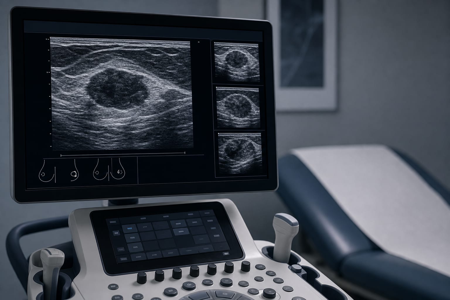
To describe the subtype-specific sonographic signatures and baseline clinicopathological features of metaplastic breast cancer (MBC) based on the World Health Organization classification.
Approach:
Study Design: Exploratory single-center retrospective cohort study including 50 women with histologically confirmed MBC.
Methods: Preoperative ultrasound features evaluated via BI-RADS criteria and compared across subtypes.
Key Findings:
Spindle cell subtype often presented with pseudo-benign sonographic phenotype (60.0% circumscribed margins, 46.7% oval/round shape).
Spindle cell tumors had significantly smaller dimensions (median 3.1 cm; p = 0.041).
Squamous cell and mixed-type tumors showed heterogeneous echogenicity (83.3% and 73.9%, respectively; p = 0.003) and complex cystic-solid components (75.0% and 56.5%, respectively; p = 0.037).
Despite a large mean primary tumor size of 4.7 cm, the overall axillary lymph node metastasis rate was 22.0%, with similar rates in tumors measuring 2–5 cm and >5 cm (26.1% and 23.8%, respectively).
Interpretation:
MBC demonstrates significant subtype-specific heterogeneity, with the spindle cell subtype posing a diagnostic challenge due to its pseudo-benign appearance.
Limitations:
Nodal events were limited and long-term survival, recurrence, and distant metastasis data were unavailable.
The study was exploratory and not confirmatory.
Conclusion:
Recognizing distinct sonographic patterns may facilitate accurate diagnosis and timely multidisciplinary management.
A-DREAM/Alliance A032101 study presented by Dana-Farber Cancer Institute's Dr. Atish Choudhury shows 41% of favorable responders to testosterone suppression plus androgen receptor pathway inhibitor remaining treatment-free with testosterone recovery at 18 months after treatment interruption.