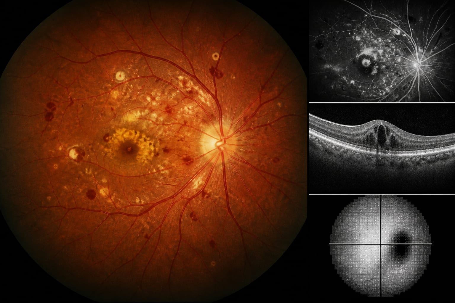
The article reviews persistent fetal vasculature (PFV), covering its developmental biology and clinical classification in the supplied excerpt.
Approach:
Developmental biology: The article describes hyaloid-vessel formation and regression, including HIF-VEGF signalling, macrophage-derived WNT7b, Norrin/FZD4/LRP5/6 signalling, and genes associated with PFV-like phenotypes.
Clinical context: The excerpt introduces Goldberg’s anterior, posterior, and combined PFV classification and describes characteristic findings, including retrolental fibrovascular plaque, ciliary-process elongation, a shallow anterior chamber, and posterior stalks, folds, or tractional detachment.
Key Findings:
PFV is uncommon and is an important cause of infantile leucocoria; it can mimic retinoblastoma.
The article describes hyaloid regression as an active process involving reduced VEGF signalling and macrophage-mediated endothelial apoptosis.
Disruption of NDP, FZD4, LRP5, or TSPAN12 can produce PFV-like features and is associated with inherited vitreoretinopathies; biallelic ATOH7 loss-of-function mutations are associated with autosomal-recessive PFV.
Goldberg’s anterior, posterior, and combined classification links clinical manifestations with the developmental continuum of PFV.
Interpretation:
The article frames PFV as a disorder involving disrupted coordination among angiogenic signalling, vascular regression, and ocular growth, with clinical manifestations spanning anterior and posterior disease.
Limitations:
The supplied excerpt ends during the clinical classification and multimodal imaging section; it does not include the remainder of the article or its detailed discussion of current treatments and therapeutic advances.
Conclusion:
The supplied excerpt connects PFV’s developmental biology with its clinical classification and characteristic ocular findings; it does not include the article’s later discussion of imaging or treatment.