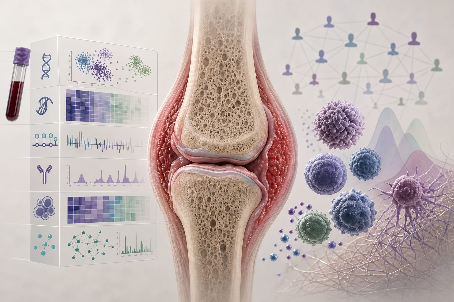
To delineate the relationship between immune cell dynamics and fibrotic remodeling in IgG4-related disease (IgG4-RD) using spatial transcriptomic analysis.
Approach:
Study Design: Spatial transcriptomic analysis of submandibular gland tissues from two patients with IgG4-RD, representing distinct fibrosis-associated tissue states.
Data Analysis: Gene expression data were normalized, z-scored, and analyzed for immune- and fibrosis-related module scores, with spatial co-localization and correlation analyses.
Key Findings:
Fibrosis-high tissue regions showed enrichment of SPP1-expressing macrophages co-localized with COL1A1 and fibroblast-rich areas.
Increased expression of PDGFB and TGF-β-related molecules correlated with fibrosis-related signatures in these regions.
Immune network analysis indicated remodeling of the immune microenvironment, with expansion of Tph-associated populations.
A preTfh-to-Tph axis was more pronounced in fibrosis-high lesions, with reduced germinal center B-cell signatures.
Interpretation:
The findings indicate a spatially coordinated relationship between immune remodeling and fibrosis in IgG4-RD, highlighting the role of SPP1 in the immune-fibrotic niche.
Limitations:
The study included a small sample size of only two patients.
Findings are based on tissue samples collected before treatment, which may not represent the disease state post-therapy.
Conclusion:
This study provides insights into fibrosis-associated tissue remodeling in IgG4-RD and may inform future strategies for disease stratification and therapeutic targeting.