CT-based habitat imaging integrated with radiomics and clinicopathology for noninvasive prediction of microvascular invasion in hepatocellular carcinoma - Summary - MDSpire
Advertisement
CT-based habitat imaging integrated with radiomics and clinicopathology for noninvasive prediction of microvascular invasion in hepatocellular carcinoma
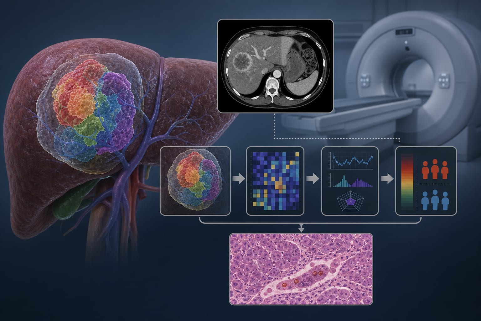
To develop and validate a CT-based habitat imaging model incorporating intratumoral microenvironment heterogeneity analysis for noninvasive preoperative prediction of microvascular invasion (MVI) in hepatocellular carcinoma (HCC).
Key Findings:
Combined model achieved an AUC of 0.862 (95% CI: 0.797–0.926) in the training set and 0.814 (95% CI: 0.710–0.918) in the validation set.
The combined model significantly outperformed individual models (P < 0.05).
Calibration curves showed good agreement (Hosmer–Lemeshow P = 0.60).
DCA indicated net benefit at thresholds of 15%–65%.
DeLong’s test confirmed higher AUC for the combined model compared to clinical and radiomics-only models.
Interpretation:
The CT-based habitat imaging model effectively quantified intratumoral heterogeneity and provided a reliable noninvasive tool for preoperative MVI risk stratification in HCC, with significant implications for clinical practice.
Limitations:
Retrospective design may introduce selection bias, potentially affecting the generalizability of the findings.
Single-center study limits generalizability of findings; further validation in larger, multicenter cohorts is needed.
Conclusion:
The study presents a novel CT-based habitat imaging model that enhances the prediction of MVI in HCC, indicating strong clinical potential and addressing limitations of existing models.