To quantitatively assess the short-term effects of RAS inhibitors on retinal microvasculature in patients with diabetic kidney disease using AI-based analysis of ultra-wide-field fundus images.
Approach:
Study Design: A prospective cohort study involving 27 patients with diabetic kidney disease (DKD) was conducted between July 2023 and September 2024.
Data Collection: Ultra-wide-field fundus images were acquired at baseline and 12 weeks after RASi therapy initiation.
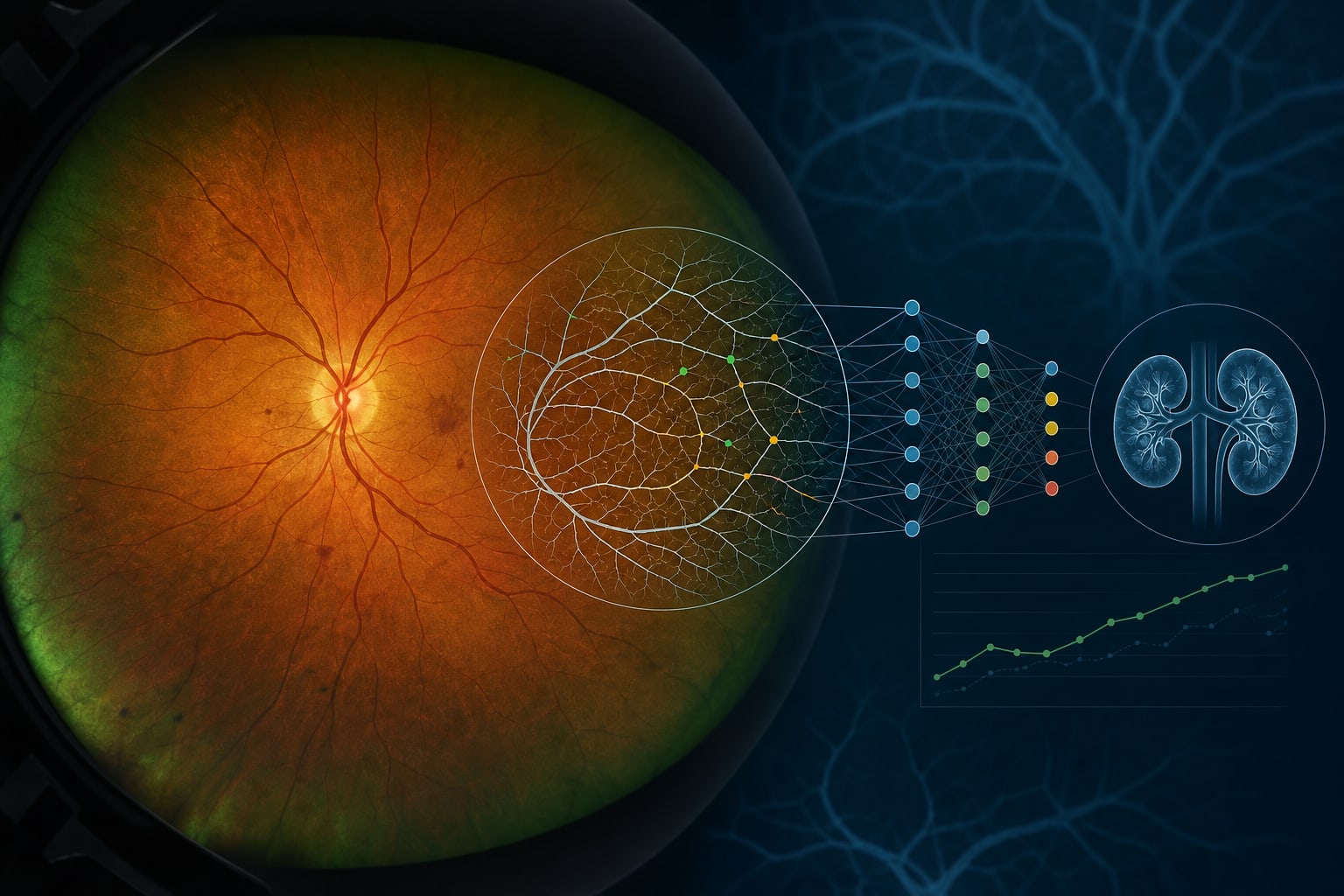
AI Analysis: A validated deep learning AI model was used to segment retinal vessels and quantify retinal microvascular parameters (RMPs).
Statistical Analysis: Pre- and post-treatment comparisons were performed using a linear mixed-effects model or Wilcoxon signed-rank test.
Key Findings:
Among the enrolled patients, 21 patients (77.8%) were male, with a mean age of 55.7 ± 14.2 years.
After 12 weeks of RASi treatment, significant decreases were observed in venous fractal dimension (Df) (adjusted p = 0.0389) and tortuosity (TORT) (adjusted p = 0.0496).
No significant changes were noted in parameters derived from the central retinal region.
Interpretation:
RASi therapy may be associated with alterations in retinal peripheral venous parameters.
Limitations:
Small sample size of 27 patients limits generalizability.
No formal sample size calculation was performed due to the exploratory nature of the study.
Conclusion:
The study indicates retinal peripheral venous alterations associated with RASi therapy.