Conexiant’s news site is now MDSpire News. Learn more
Advertisement
Lumbar disc herniation may mask coral reef aorta
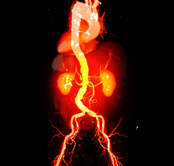
Serial imaging documented 6 years of progressive aortic calcification, culminating in near occlusion before open surgical reconstruction restored blood flow.
To describe progressive coral reef aorta causing severe lower-extremity ischemia in a patient whose symptoms were initially attributed to lumbar disc herniation.
Approach:
Case and assessment: Researchers reported a 50-year-old man with smoking, hypertension, and hyperlipidemia, reviewing six years of CT scans alongside clinical examinations and ankle-brachial index (ABI) measurements.
Treatment and follow-up: Because extensive aortic calcification made endovascular treatment unsuitable, the patient underwent open aortic graft replacement with inferior mesenteric artery reconstruction. Clinical status, ABI, and contrast-enhanced CT were assessed through one year.
Key Findings:
Bilateral leg numbness had been attributed to lumbar disc herniation four years earlier; the patient later developed claudication and severe rest pain.
ABI at presentation was 0.49 in both legs, compared with 0.99 on the right and 1.07 on the left six years earlier.
CT showed dense calcified plaques extending more than 4 cm along the infrarenal aorta and causing near occlusion; earlier scans showed progressive calcification.
After surgery, symptoms resolved and ABI improved to 1.13 on the right and 1.19 on the left by postoperative day four.
At one year, the patient remained asymptomatic, ABI was 1.07 bilaterally, and CT showed patent graft and reconstructed artery.
Interpretation:
The authors noted that coexisting lumbar disc herniation may have masked the vascular disorder. They stated that coral reef aorta should be considered in unexplained progressive bilateral lower-extremity ischemia and that treatment selection should reflect anatomy and patient risk.
Limitations:
The single-patient report cannot establish comparative effectiveness of open surgery versus endovascular treatment.
The extent to which the initial diagnosis delayed recognition of vascular disease could not be determined definitively.
One-year follow-up does not establish longer-term graft durability or risk of recurrent vascular complications.
Conclusion:
Serial CT documented progressive aortic calcification and informed treatment selection in this case. The authors reported no conflicts of interest or funding.
Review identifies recurring sex-based differences across specialties, including lower use of intensive and guideline-compliant treatment among female patients.