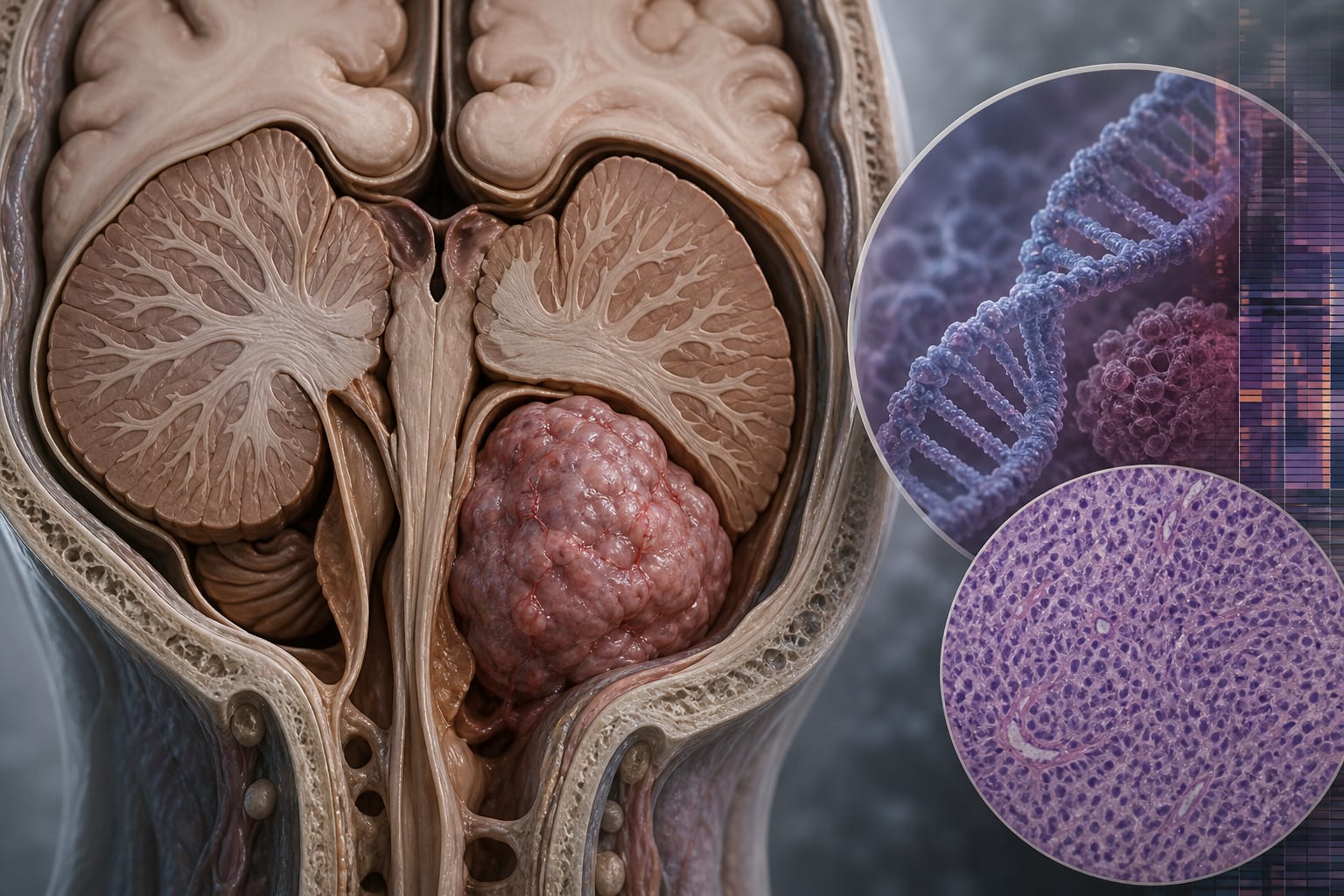
Posterior fossa ependymoma: a comprehensive review of molecular classification, management guidelines, and clinical outcomes (Part I of ependymomas across compartments) - Takeaways - MDSpire
Coming Soon: Introducing MDSpire News. Learn more
Conexiant’s news site is now MDSpire News. Learn more

Ependymomas in the Posterior Fossa: An In-Depth Analysis of Molecular Classification, Treatment Protocols, and Clinical Outcomes (Part I on Ependymomas in Various Locations)

  • By

  • George W. Koutsouras

  • Francisco Rivera

  • Anthony M. Price

  • Derek S. Tsang

  • Adam Esbenshade

  • Vijay Ramaswamy

  • Peter B. Dirks

  • Michael C. Dewan

  • September 22, 2026

Share

  • 1

    Ependymal tumors account for about 5% of pediatric CNS tumors, with two-thirds originating in the posterior fossa.

  • 2

    Molecular profiling has shifted WHO classification of ependymomas from histologic to integrated molecular entities, including PFA and PFB.

  • 3

    PFA tumors are linked to poorer clinical outcomes due to epigenetic changes, while PFB tumors retain PRC2 regulation and show higher somatic aneuploidy.

  • 4

    MRI is essential for diagnosing posterior fossa ependymomas, revealing differences in imaging phenotypes between PFA and PFB tumors.

  • 5

    Surgical management aims for gross total resection, which significantly improves survival outcomes compared to subtotal resection.

Original Source(s)

Related Content