A 30-year-old woman had multiple rim-enhancing brain lesions on routine MRI despite no initial fever, pulmonary symptoms, or neurologic abnormalities.
2
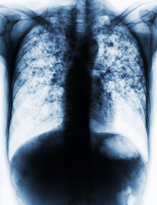
The MRI findings suggested toxoplasmosis or neurocysticercosis, but chest CT showed bilateral 1- to 3-mm pulmonary nodules consistent with miliary tuberculosis.
3
Sputum PCR detected Mycobacterium tuberculosis complex DNA, whereas sputum cultures and CSF tuberculosis PCR were negative and CSF analysis was unremarkable.
4
The researchers diagnosed probable parenchymal CNS tuberculosis with miliary pulmonary tuberculosis and treated the patient with antituberculosis drugs and adjunctive corticosteroids.
5
After 12 months of therapy, brain lesions regressed, and complete clinical and radiologic remission was documented 12 months after treatment.