Intratumoral and peritumoral MRI habitat imaging for differentiating stage IA endometrial cancer from benign endometrial lesions: a multicenter study - Takeaways - MDSpire

Intratumoral and peritumoral MRI habitat imaging for differentiating stage IA endometrial cancer from benign endometrial lesions: a multicenter study

  • By

  • Yunzhu Wu

  • Xianhong Wang

  • Cheng Deng

  • Qiu Bi

  • Yang Song

  • Shenghong Ju

  • Yang Liu

  • Conghui Ai

  • Jing Yang

  • July 9, 2026

  • 0 min

Share

  • 1

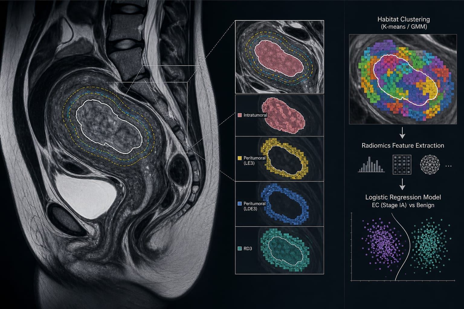
    This study evaluated multiparametric MRI-based radiomics models to differentiate stage IA endometrial cancer from benign lesions.

  • 2

    A total of 787 patients were included, with tumor regions manually delineated on MRI and peritumoral ROIs generated automatically.

  • 3

    Age and vaginal bleeding were identified as independent clinical predictors of stage IA endometrial cancer.

  • 4

    The combined model of clinical predictors and habitat features achieved the highest diagnostic performance with a peak AUC of 0.921.

  • 5

    K-means clustering demonstrated superior robustness compared to Gaussian Mixture Model in the analysis of peritumoral features.

Original Source(s)

Related Content