Adenomatoid tumors of the uterus: magnetic resonance imaging features with clinical and histopathologic correlation - Takeaways - MDSpire
Coming Soon: Introducing MDSpire News. Learn more
Conexiant’s news site is now MDSpire News. Learn more

Magnetic Resonance Imaging Characteristics of Uterine Adenomatoid Tumors: A Study of Clinical and Histopathological Correlations

  • By

  • Changhuan Chen

  • Yudian Huang

  • Ping Chen

  • Caixia Zheng

  • Chengsheng Wang

  • Mingsi Yang

  • September 8, 2026

Share

  • 1

    Uterine adenomatoid tumors (ATs) are uncommon benign neoplasms arising from mesothelial tissue, often discovered incidentally during histopathological examination.

  • 2

    The study analyzed 16 surgically resected uterine ATs, revealing a median patient age of 51 years and coexistence with uterine fibroids in 8 cases.

  • 3

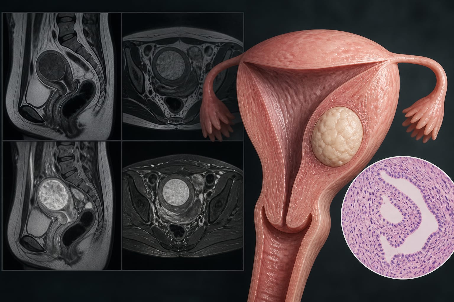
    MRI characteristics showed that ATs typically appear as solid masses with well-defined margins, lacking a discernible capsule, and may exhibit cystic degeneration.

  • 4

    On T1-weighted imaging, solid components of ATs are isointense, while T2-weighted imaging predominantly shows them as hypointense with possible 'target sign' features.

  • 5

    Histopathologically, uterine ATs consist of gland-like spaces among smooth muscle and fibrous tissue, correlating closely with their MRI findings.

Original Source(s)

Related Content