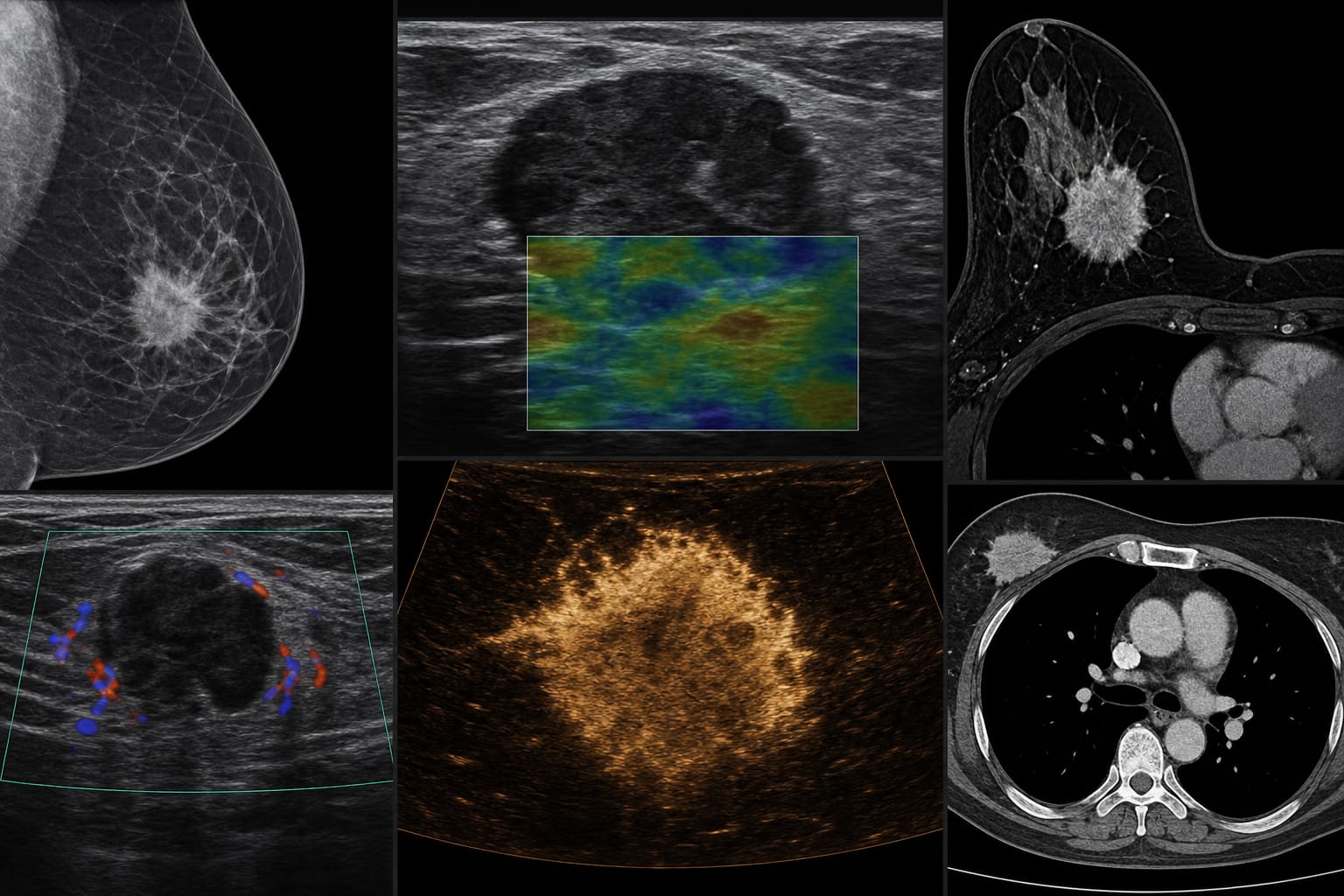
Multimodal imaging features of primary breast epithelial–myoepithelial carcinoma: a case report and literature review
-
By
-
Peilin Sha
-
Yunhao Luo
-
Xiangzhu Wang
-
Yanhong An
-
Jia Jia
-
Xue Shi
-
Xin Li
-
Mengke Xu
-
Huiyu Ge
-
Zexing Yu
-
July 1, 2026
-
0 min