Pulmonary Kaposi Sarcoma in Advanced HIV With Flame-Like Opacities on Chest CT
-
October 6, 2026
-
1
A 61-year-old man with advanced HIV, progressive dyspnea, productive cough, and violaceous skin lesions was ultimately diagnosed with pulmonary Kaposi sarcoma.
-
2
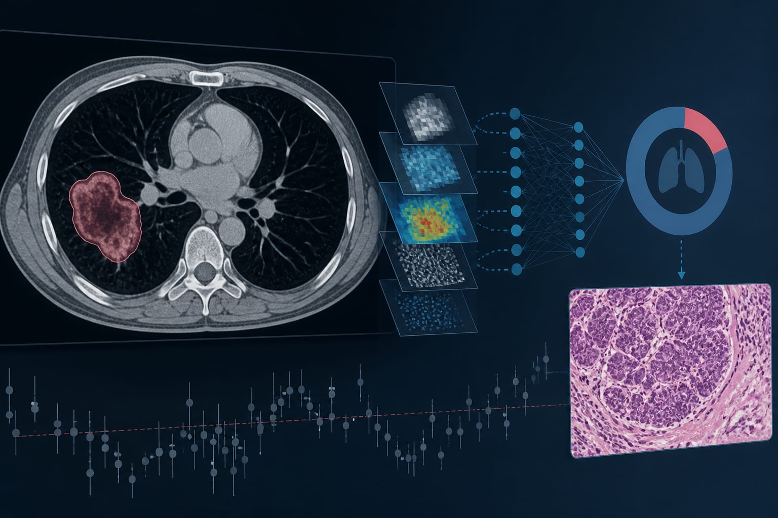
Chest CT showed bilateral, asymmetric, flame-shaped nodular opacities along the peri-bronchovascular regions, predominantly on the left.
-
3
The initial scant left lung core biopsy suggested adenocarcinoma but was inconsistent with the clinical and radiographic findings.
-
4
Repeat CT-guided samples from the left lower lobe and lingula showed spindle-cell vascular proliferation and human herpesvirus 8 latent nuclear antigen-1 positivity, confirming Kaposi sarcoma.
-
5
The case authors recommend repeat tissue sampling with human herpesvirus 8 immunohistochemistry when pathology conflicts with compelling clinical and imaging findings.
Based on findings from:
Pulmonary Kaposi Sarcoma Presenting With Flame-Shaped Computed Tomography Opacities in Advanced HIV
Annals Of Internal Medicine: Clinical Cases, 2026.
https://www.acpjournals.org/doi/10.7326/aimcc.2026.0557
This content is an AI-generated, fully rewritten summary based on a published scholarly article. It does not reproduce the original text and is not a substitute for the original publication. Readers are encouraged to consult the source for full context, data, and methodology.