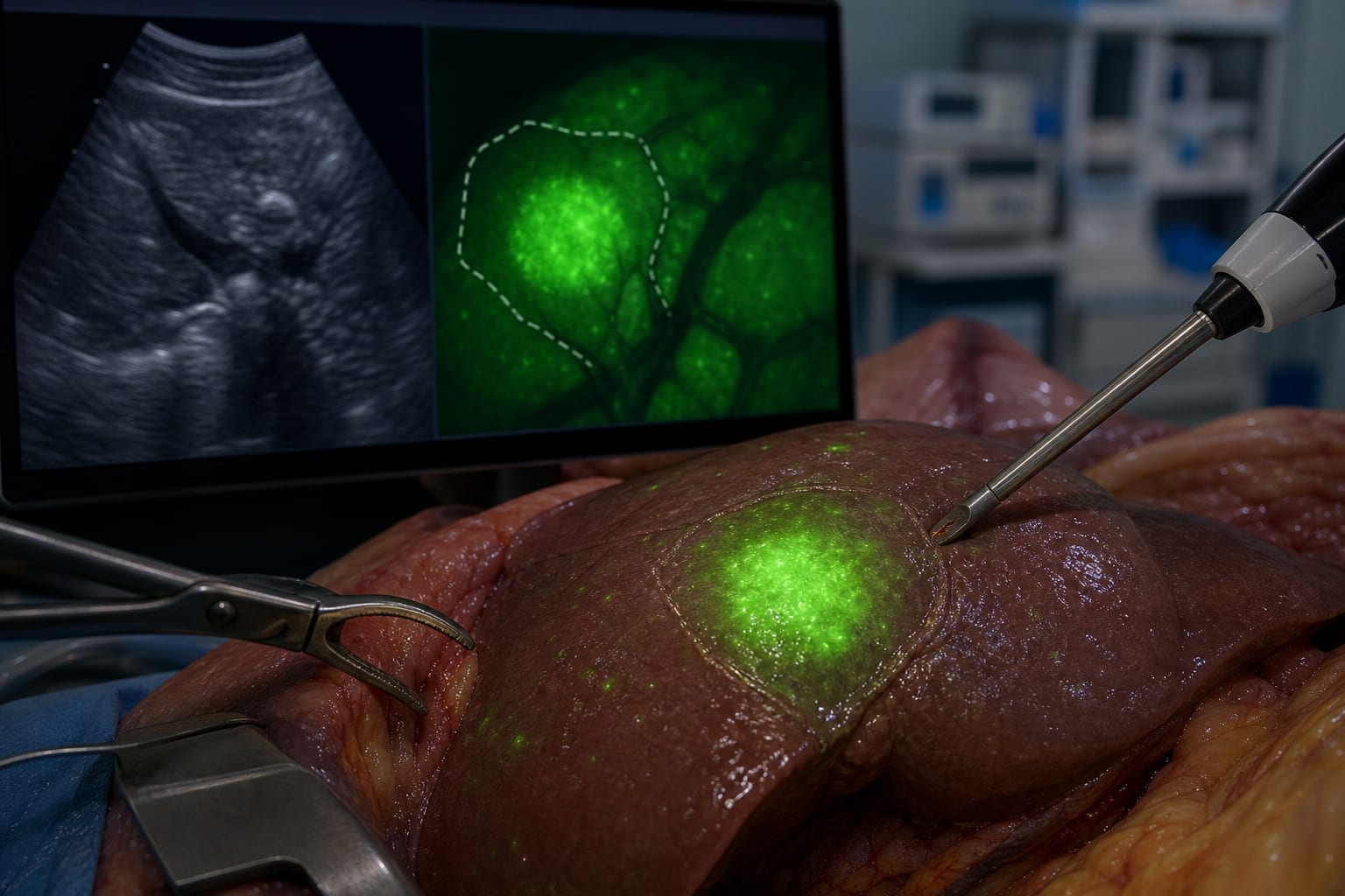
Case Report: Indocyanine green fluorescence imaging in complex focal nodular hyperplasia resection: report of two cases
-
By
-
Fangkai Du
-
Xie Song
-
Wentao Wang
-
Huizhong Shi
-
Zhengjian Wang
-
Chaoqun Ma
-
Qingqiang Ni
-
Shunzhen Zheng
-
Hong Chang
-
June 15, 2026
-
0 min