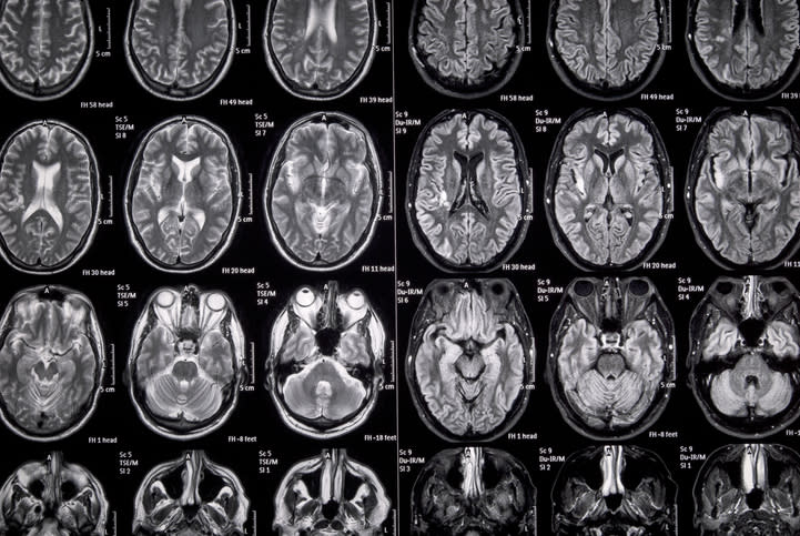
Meningococcal Abscess Detected by MRI
MRI identifies rare intracranial complication following normal computed tomography
-
By
-
Julie Greenbaum
-
March 27, 2026
-
-
1
An 11-year-old boy with meningococcal meningitis developed a cerebellar abscess detectable only by MRI, despite normal CT findings.
-
2
Cerebrospinal fluid analysis revealed elevated white blood cells and confirmed Neisseria meningitidis as the causative agent.
-
3
The patient's clinical condition improved with antimicrobial therapy, leading to extubation and transfer out of intensive care by day 5.
-
4
MRI demonstrated the evolution of the cerebellar abscess, which resolved without surgical intervention, guided by multidisciplinary consensus.
-
5
The case highlights the diagnostic superiority of MRI over CT in detecting early parenchymal infections in atypical meningitis presentations.