Size and location of intracranial hemorrhage in acute stroke presentations: correlation with baseline NIHSS and relevance to direct-to-angiography workflows - Takeaways - MDSpire
Coming Soon: Introducing MDSpire News. Learn more
Conexiant’s news site is now MDSpire News. Learn more

Assessment of Intracranial Hemorrhage Size and Location in Acute Stroke Cases: Its Relationship with Initial NIHSS Scores and Implications for Direct-to-Angiography Protocols

  • By

  • Anh Nguyen

  • Mira Katan

  • Raphael Guzman

  • Jehuda Soleman

  • Christian Nickel

  • Marios-Nikos Psychogios

  • Alex Brehm

  • September 8, 2026

Share

  • 1

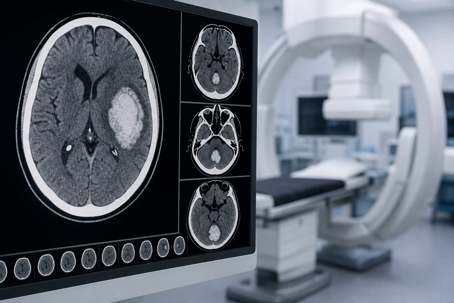
    Intraparenchymal hemorrhage accounted for 89% of cases in the study, with median volume measured at 18 mL.

  • 2

    Volume of intracranial hemorrhage significantly correlated with NIHSS scores, indicating a relationship between size and stroke severity.

  • 3

    Patients with NIHSS scores ≥7 had larger hemorrhages compared to those with scores <7, with median volumes of 38 mL and 6 mL, respectively.

  • 4

    Lobar hemorrhages exhibited the greatest volumes, while infratentorial hemorrhages, particularly in the brainstem, caused severe deficits despite smaller volumes.

  • 5

    The study highlights the challenges in detecting small-volume infratentorial hemorrhages on FDCT, emphasizing the need for improved imaging quality.

Original Source(s)

Related Content