Functional ultrasound assessment of cerebral blood flow and brain connectivity in a pilocarpine-induced acute epileptic seizures in mice - Takeaways - MDSpire
Coming Soon: Introducing MDSpire News. Learn more
Conexiant’s news site is now MDSpire News. Learn more

Evaluation of Cerebral Blood Flow and Brain Connectivity Using Functional Ultrasound in Mice with Pilocarpine-Induced Acute Seizures

  • By

  • Yao Liu

  • Chang Xu

  • Wenqian Ma

  • Dan Zhao

  • Baocong Yu

  • Rui Zhang

  • Haiyue Huang

  • Jianguo Niu

  • Ling He

  • Yujun Wen

  • June 15, 2026

Share

  • 1

    Temporal lobe epilepsy (TLE) is a prevalent form of epilepsy with no effective curative treatments currently available.

  • 2

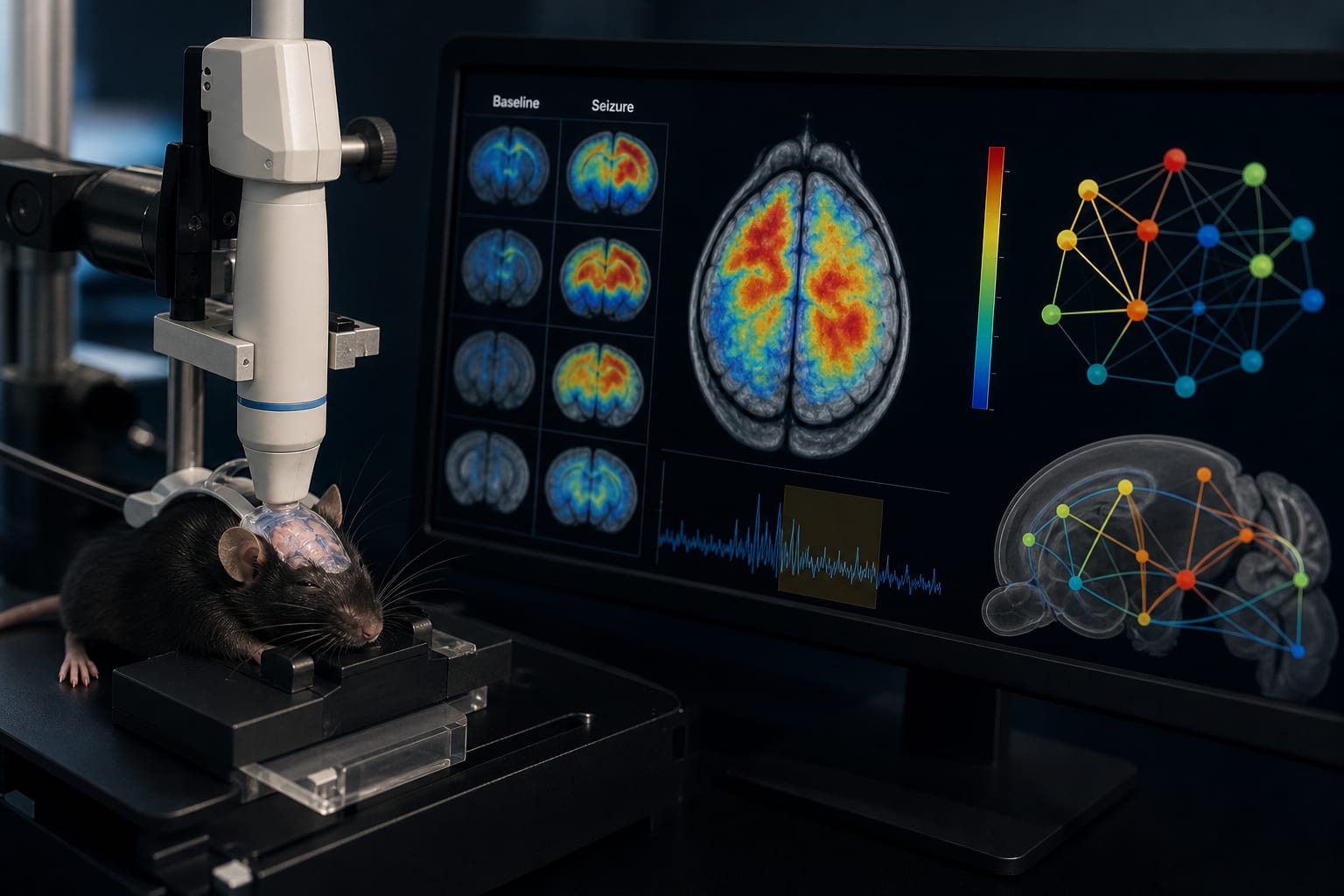
    Functional ultrasound imaging (fUS) was utilized to assess changes in cerebral blood volume (CBV) during acute seizure states in mice.

  • 3

    Most brain regions showed changes in relative CBV, with all but two regions exhibiting an initial increase followed by a decrease.

  • 4

    The reticular nucleus of the thalamus (RT) exhibited earlier changes in rCBV compared to other brain regions.

  • 5

    Overall brain functional network connectivity underwent significant modifications during the acute seizure state in TLE mice.

Original Source(s)

Related Content